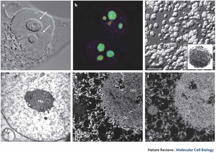
Nucleolus is the largest and the first membrane-less organelle which was identified. It is composed of a fibrillar centre (FC), dense fibrillar component (DFC) and granular component (GC). The nucleolus is a dynamic structure that disassembles during mitosis and reassembles after cell division. The nucleolus is responsible for ribosome biogenesis in eucaryotic cells, which are essential macromolecular machines responsible for synthesizing all proteins required by the cell. The nucleoli are now known to act as sensors and regulators of cellular responses, such as apoptosis, DNA damage response, epigenetic modification, genome stability, proliferation, senescence and signal transduction (reviewed in 34358306, 31405125, 17519961).

Figure 1: Different imaging techniques can be used to identify distinct aspects of nucleolar morphology and composition. The fibrillar centre (FC), dense fibrillar component (DFC) and granular component (GC) regions can be individually visualized using transmission electron microscopy (EM) and specifically labelled by fluorescence microscopy using reporter proteins fused to fluorescent protein tags. The dense shell of heterochromatin that surrounds nucleoli can be identified by scanning EM of either intact cells or isolated nucleoli, or labelled by 4′,6-diamidino-2-phenylindole (DAPI) in the fluorescence microscope. a | Differential interference contrast (DIC) image of a HeLa cell showing prominent nucleoli within the nucleus (indicated by arrows). b | Immunofluorescence labelling of a HeLa cell with antibodies that are specific for proteins enriched in the GC (B23; shown in green), the DFC (fibrillarin; shown in red) or the FC (RNA polymerase I subunit RPA39; shown in blue). c | DIC image of nucleoli purified from HeLa cells. The inset shows a scanning EM image of a purified HeLa nucleolus. d | Uranyl-acetate-stained cell section showing a characteristic image of a nucleus with a nucleolus (Nu) imaged by transmission EM. e,f | Ultrastructural analysis of nucleoli by electron spectroscopic imaging (ESI). A nuclear region of interest that contains the nucleolus with phosphorus (e) and nitrogen (f) enriched images that reveal nucleic-acid-based and protein-based components, respectively (Nature Reviews Molecular Cell Biology (Nat Rev Mol Cell Biol) ISSN 1471-0080 (online) ISSN 1471-0072 (print) (17519961).
¶ Contents
Formation, Composition & dynamics (Assembly and disassembly)
Relation to human diseases
Proteome
Formation, Composition & dynamics (Assembly and disassembly)
The nucleolus is a key organelle that controls the synthesis and assembly of ribosomal subunits. The nucleolus is a multilayered biomolecular condensate made up of three phase separated layers: the fibrillar center (where ribosomal DNA is translated into rRNA), the dense fibrillar component (which is responsible for rRNA processing), and the granular component. The nucleolar structure is formed by changes in surface tension between the different liquid phases that result from their macromolecular components. The nucleolus's liquid droplet-like characteristics enable it to play a variety of dynamic functions. The liquid droplet generation property explains a variety of nucleolar functions by isolating the nucleolus from the nucleoplasm despite the absence of a membrane (detailed reviews: 31405125, 2873929, 34358306).
28600324 identified NPM2 as the main component which is necessary for structural integrity of the oocyte nucleolus and showed that oocyte nucleolus can be reconstituted only by the expression of NPM2.
Relation to human diseases
Ribosomes are essential for protein production, and hence for cellular survival, growth and proliferation. Ribosome biogenesis begins in the nucleolus and therefore nucleolus could cause various cellular problems when impaired.
31026227, 33120992, 30650663 reviewed impaired ribosome biogenesis and its relation to aging and cancer.
In other circumstances, independent of its functions in ribosome synthesis, the nucleolus has emerged as a critical controller of many cellular processes that are essential to normal cell homeostasis and the target of dysregulation in many human disorders.
29603296 reviewed novel roles of nucleolus in human diseases independent of ribosome biogenesis.
32106410 addressed new insights into the physical and molecular mechanisms that control the architecture and diverse functions of the nucleolus, and their failure in disease.
26427048 reviewed the role of the nucleolus in retinal development as well as in neurodegeneration.
30634859 summarized recent advances on nucleolar functions in health and disease.
31338556 addresses the central role of the nucleolus in carcinogenesis and cancer progression.
27264829 discusses nucleolar stress in Parkinson’s disease.
Proteome
Purification and extensive proteomic analyses of nucleoli have contributed to understanding of their multiple biological functions. The following references provide useful data on the proteomic of nucleoli.
15635413 used mass-spectrometry-based organellar proteomics and stable isotope labelling to quantitatively analyze the proteome dynamics of human nucleoli. The data establish a quantitative proteomic approach for the temporal characterization of protein flux through cellular organelles and demonstrate that the nucleolar proteome changes significantly over time in response to changes in cellular growth conditions.
2014684 used the Jurkat T-cell line and a reproducible organellar proteomic approach and identified 872 nucleolar proteins.
20970743 used LC-MS/MS to analyze the proteomic composition of the bovine nucleoli and identified 311 proteins in the bovine nucleoli, which contained 22 proteins previously not identified in the proteomic analysis of human nucleoli.
26980300 analyzed the proteome of the nucleus and nucleolus of Arabidopsis thaliana and identified 1602 proteins in the nucleolar and 2544 proteins in the nuclear fraction with an overlap of 1429 proteins.
References
Yoneda M, Nakagawa T, Hattori N, Ito T. The nucleolus from a liquid droplet perspective. J Biochem. 2021 Oct 11;170(2):153-162. doi: 10.1093/jb/mvab090. PMID: 34358306.
Shiroma K, Suma K, Kawai Y, Kaneko H, Miyawaki F, Imanishi K, Torii S, Mukai F, Suda Y. [Mitral valve surgery using combined superior-transseptal approach to the left atrium]. Kyobu Geka. 1992 Nov;45(12):1071-4. Japanese. PMID: 1405125.
Boisvert FM, van Koningsbruggen S, Navascués J, Lamond AI. The multifunctional nucleolus. Nat Rev Mol Cell Biol. 2007 Jul;8(7):574-85. doi: 10.1038/nrm2184. PMID: 17519961.
Correll CC, Bartek J, Dundr M. The Nucleolus: A Multiphase Condensate Balancing Ribosome Synthesis and Translational Capacity in Health, Aging and Ribosomopathies. Cells. 2019 Aug 10;8(8):869. doi: 10.3390/cells8080869. PMID: 31405125; PMCID: PMC6721831.
Goodman MN, Lowenstein JM. The purine nucleotide cycle. Studies of ammonia production by skeletal muscle in situ and in perfused preparations. J Biol Chem. 1977 Jul 25;252(14):5054-60. PMID: 873929.
Taylor HW, Olson LD. Chronologic study of the T-virus in chicks. I. Development of lesions. Avian Dis. 1973 Oct-Dec;17(4):782-94. PMID: 4358306.
Ogushi S, Yamagata K, Obuse C, Furuta K, Wakayama T, Matzuk MM, Saitou M. Reconstitution of the oocyte nucleolus in mice through a single nucleolar protein, NPM2. J Cell Sci. 2017 Jul 15;130(14):2416-2429. doi: 10.1242/jcs.195875. Epub 2017 Jun 9. PMID: 28600324; PMCID: PMC6139372.
Turi Z, Lacey M, Mistrik M, Moudry P. Impaired ribosome biogenesis: mechanisms and relevance to cancer and aging. Aging (Albany NY). 2019 Apr 26;11(8):2512-2540. doi: 10.18632/aging.101922. PMID: 31026227; PMCID: PMC6520011.
Nait Slimane S, Marcel V, Fenouil T, Catez F, Saurin JC, Bouvet P, Diaz JJ, Mertani HC. Ribosome Biogenesis Alterations in Colorectal Cancer. Cells. 2020 Oct 27;9(11):2361. doi: 10.3390/cells9112361. PMID: 33120992; PMCID: PMC7693311.
Penzo M, Montanaro L, Treré D, Derenzini M. The Ribosome Biogenesis-Cancer Connection. Cells. 2019 Jan 15;8(1):55. doi: 10.3390/cells8010055. PMID: 30650663; PMCID: PMC6356843.
Núñez Villacís L, Wong MS, Ferguson LL, Hein N, George AJ, Hannan KM. New Roles for the Nucleolus in Health and Disease. Bioessays. 2018 May;40(5):e1700233. doi: 10.1002/bies.201700233. Epub 2018 Mar 30. PMID: 29603296.
Stochaj U, Weber SC. Nucleolar Organization and Functions in Health and Disease. Cells. 2020 Feb 25;9(3):526. doi: 10.3390/cells9030526. PMID: 32106410; PMCID: PMC7140423.
Sia PI, Wood JP, Chidlow G, Sharma S, Craig J, Casson RJ. Role of the nucleolus in neurodegenerative diseases with particular reference to the retina: a review. Clin Exp Ophthalmol. 2016 Apr;44(3):188-95. doi: 10.1111/ceo.12661. Epub 2016 Feb 5. PMID: 26427048.
Bahadori M, Azizi MH, Dabiri S. Recent Advances on Nucleolar Functions in Health and Disease. Arch Iran Med. 2018 Dec 1;21(12):600-607. PMID: 30634859.
Weeks SE, Metge BJ, Samant RS. The nucleolus: a central response hub for the stressors that drive cancer progression. Cell Mol Life Sci. 2019 Nov;76(22):4511-4524. doi: 10.1007/s00018-019-03231-0. Epub 2019 Jul 23. PMID: 31338556; PMCID: PMC6841648.
Zhou Q, Chen Y, Wei Q, Shang H. [Parkinson's disease and nucleolar stress]. Zhonghua Yi Xue Yi Chuan Xue Za Zhi. 2016 Jun;33(3):392-5. Chinese. doi: 10.3760/cma.j.issn.1003-9406.2016.03.026. PMID: 27264829.
Andersen JS, Lam YW, Leung AK, Ong SE, Lyon CE, Lamond AI, Mann M. Nucleolar proteome dynamics. Nature. 2005 Jan 6;433(7021):77-83. doi: 10.1038/nature03207. PMID: 15635413.
Slisow W, Möhner M. Metastazirovanie raka priamoĭ kishki [The metastasis of rectal cancer]. Vopr Onkol. 1991;37(1):76-80. Russian. PMID: 2014684.
Patel AK, Olson D, Tikoo SK. Proteomic analysis of bovine nucleolus. Genomics Proteomics Bioinformatics. 2010 Sep;8(3):145-58. doi: 10.1016/S1672-0229(10)60017-4. PMID: 20970743; PMCID: PMC5054126.
Palm D, Simm S, Darm K, Weis BL, Ruprecht M, Schleiff E, Scharf C. Proteome distribution between nucleoplasm and nucleolus and its relation to ribosome biogenesis in Arabidopsis thaliana. RNA Biol. 2016;13(4):441-54. doi: 10.1080/15476286.2016.1154252. Epub 2016 Mar 16. PMID: 26980300; PMCID: PMC5038169.